Correlating Fundus Flavoprotein Fluorescence with Structural Changes in Geographic Atrophy
 Britney A. Naolhu, Sairi Zhang, Jeong W. Pak, Rick Voland, Amitha Domalpally, Collin Rich, Mihai Mititelu
Abstract
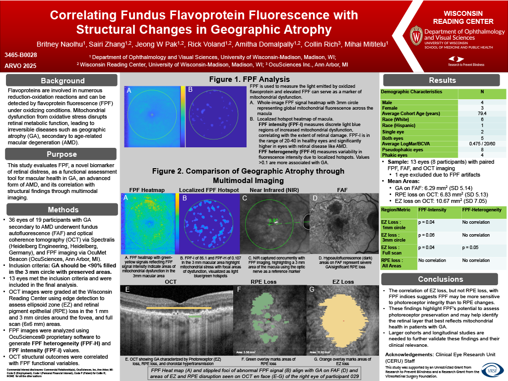
Purpose: Mitochondrial dysfunction from oxidative stress disrupts retinal metabolic function, leading to diseases such as age-related macular degeneration (AMD). This study evaluates flavoprotein fluorescence (FPF), a novel biomarker of retinal distress, as a functional assessment tool for macular health in geographic atrophy (GA), an advanced form of AMD, and its correlation with structural findings through multimodal imaging.
Methods: 36 eyes of 19 participants with GA secondary to AMD underwent fundus autofluorescence (FAF) and optical coherence tomography (OCT) via Spectralis (Heidelberg Engineering, Heidelberg, Germany), and FPF imaging via OcuMet Beacon (OcuSciences, Ann Arbor, MI). OCT images were graded at the Wisconsin Reading Center using edge detection to assess ellipsoid zone (EZ) and retinal pigment epithelial (RPE) loss in the 1 mm and 3 mm circles around the fovea, and full scan (6×6 mm) areas. FPF images were analyzed using OcuSciences© proprietary software to generate FPF heterogeneity (FPF-H) and FPF intensity (FPF-I) values. 13 eyes with GA confined to the central 3 mm subfield were included in the final analysis. OCT structural outcomes were correlated with FPF functional variables.
Results: Of the 13 eyes (8 participants) with paired FPF, FAF and OCT imaging, 1 eye was excluded due to artifacts in FPF imaging. Mean area of GA on FAF was 6.29 mm2 (SD 5.14), and mean area of RPE and EZ loss on OCT was 6.83 mm2 (SD 5.13) and 10.67 mm2 (SD 7.05), respectively. EZ loss in the 1 mm circle significantly correlated with FPF-I (p = 0.04) and approached significance within the 3 mm circle (p = 0.06). FPF-H did not have a significant correlation with either 1 mm or 3 mm circles. For the full scan, EZ loss correlated with both FPF-I (p = 0.04) and FPF-H (p = 0.05). In contrast, RPE loss showed no correlation with FPF-I or FPF-H across all areas.
Conclusions: The correlation of EZ loss, but not RPE loss, with FPF indices suggests FPF may be more sensitive to photoreceptor integrity than to RPE changes. These findings highlight FPF’s potential to assess photoreceptor preservation and may help identify the retinal layer that best reflects mitochondrial health in patients with GA. Larger cohorts and longitudinal studies are needed to further validate these findings and their clinical relevance.