Prediction of Geographic Atrophy Enlargement using Various Deep Learning Approaches (2023)
 Amitha Domalpally, Robert Slater, Mark Banghart, Roomasa Channa, Donald S. Fong, Barbara Blodi
Abstract
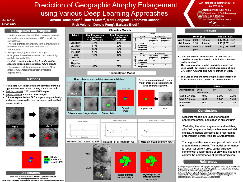
Purpose: Fundus autofluorescence (FAF) imaging is used to monitor geographic atrophy (GA) growth inclinical trials. There is significant variability in the growth rate of GA with studies reporting between 0.5 – 2.6mm/year. Multiple imaging risk factors for rapid enlargement have been studied but do not fully explain thevariability. The purpose of this project is to use AI to predict GA enlargement using various approaches.
Methods: FAF images of 338 eyes taken one year apart were included. Both visits were evaluated by expertgraders to produce area measurement and an annotation. The AI model was trained on 121 registered pairedvisits (67 subjects) and validated on 48 (23 subjects). 3 different models were trained: (1) to classify growth asfast / slow based on a cut off 1.6 mm/ year (2) to predict the area of GA at visit 2 when provided area at visit 1(3) to predict the GA mask at year 2 when provided with a mask for year 1. Validation metrics includedcomparison of grader measurement with AI predicted GA area at visit 2. Dice coefficient was used to comparethe similarity between grader annotation and AI mask.
Results: Mean area of visit 1 was 6.76mm and 8.23mm at visit 2 as measured by graders. With model 1, theaccuracy of predicting fast vs slow growth was 69% (F1 score 71%). With model 2, AI predicted area at visit 2was 8.23 mm (mean difference 0.00 mm2 (95% CI -1.72,1.72) R 0.91). The Dice coefficient between graderannotation and AI mask was 0.53.
Conclusions: AI based prediction models can be used to enrich clinical trial population with GA with fastergrowth rate. AI models for prediction of future areas and classification of fast /slow responders seem to performbetter than providing masks representing future GA. External validation on independent datasets is required to implement these models in prospective trials.