Comparison of GA measurements among ORC, OCT, and FAF imaging
 Justin Bitner, Jeong W. Pak, Jessica Berg, Rick Voland, Ronald Danis, David Bingaman, Yijun Huang, Amitha Domalpally
Abstract
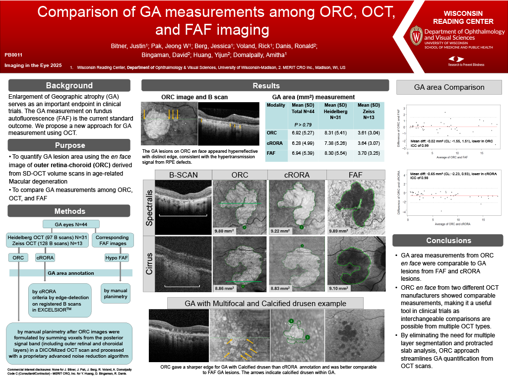
Purpose: To quantify geographic atrophy (GA) lesion area using the en face image of outer retina-choroid (ORC) derived from spectral domain-optical coherence tomography (SD-OCT) volume scans and to compare GA measurements among ORC, OCT, and fundus autofluorescence (FAF) in age-related macular degeneration.
Methods: Eyes with GA from 31 Spectralis OCTs, 13 Cirrus OCTs, and the corresponding FAF images were included. The GA areas were independently measured by manual planimetry from ORC and FAF images. ORC images were formulated by summing voxels from the posterior signal band (including outer retinal and choroidal layers) in a DICOMized OCT scan and processed with a proprietary advanced noise reduction algorithm. In addition, GA area was delineated with complete retinal pigment epithelial and outer retinal atrophy (cRORA) criteria by edge-detection on registered B scans in EXCELSIORTM. The GA lesions on ORC appeared hyperreflective with distinct edge, consistent with the hypertransmission signal from retinal pigment epithelial (RPE) defects (Figure 1).
Results: Of total 44 eyes, mean (mm2) (SD) GA area was 6.92(5.27) with ORC, 6.28(4.99) with cRORA criteria, and 6.94(5.39) with FAF. The ORC measurements closely matched FAF with a mean difference of -0.02mm2 and ICC of 0.99. The cRORA were also similar to ORC, although systematically underestimated, with a mean difference of -0.65mm2 and ICC of 0.98. When Spectralis and Cirrus OCT were analyzed separately for ORC vs FAF and for OCT vs ORC, the mean difference was -0.01mm2(ICC=0.99) and 0.04mm2(ICC=1), respectively from Cirrus, while 0.01mm2(ICC=0.99) and -0.93 mm2(ICC=0.97), respectively from Spectralis.
Conclusions: GA area measurements from ORC were comparable to GA lesions from FAF and cRORA. In addition, ORC from two different OCT manufacturers showed comparable measurements, making it a useful vendor agnostic imaging endpoint in clinical trials as interchangeable comparisons are possible from multiple OCT types. By eliminating the need for segmentation and protracted slab analysis, this approach streamlines GA quantification from OCT.