Real World Validation of Optos AI Algorithm for GA area Measurement
Reeva Faisal, Caleb Pacheco, Robert Slater, Mohammed S. Younis, Jomol Matthew, Marine Nalbandyan, Amitha Domalpally, Roomasa Channa
Abstract
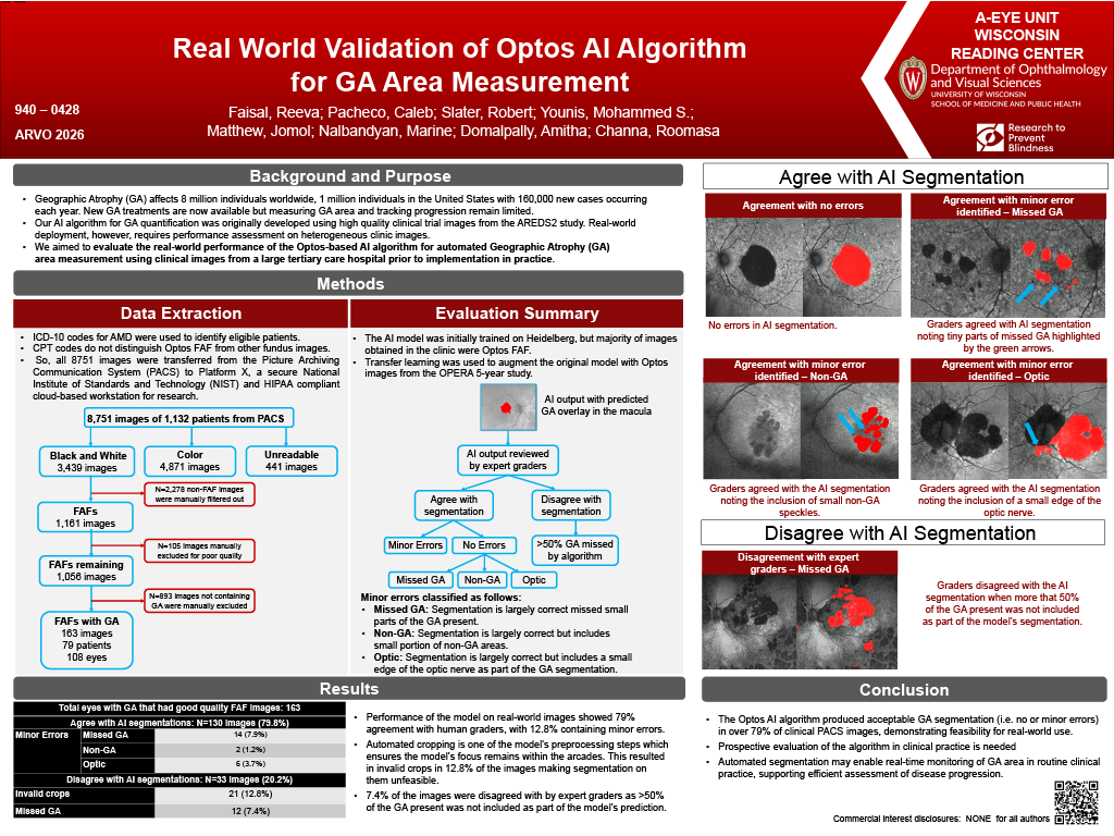
Purpose: Optos ultrawide field imaging is widely used in routine care to monitor Geographic Atrophy (GA) progression due to ease of imaging and comfort to patients. Our AI algorithm for GA quantification was originally developed and validated using high quality clinical trial images from the AREDS2 OPERA study and showed high performance metrics. Real-world deployment, however, requires performance assessment on heterogeneous clinic images. This study evaluates the real-world performance of the Optos-based AI algorithm for automated GA measurement using clinical images from a large tertiary care hospital prior to implementation in practice.
Methods: Using ICD-10 codes for GA, UW Clinical Research Data Services identified eligible patients and transferred de-identified images from the Picture Archiving and Communication System (PACS) to Platform X, a secure NIST and HIPAA compliant cloud-based workstation for research. Because CPT codes do not distinguish Optos FAF from other fundus images, all images meeting ICD-10 criteria for GA were transferred. After identifying Optos images, the validated AI model was deployed within Platform X, and automated GA segmentations were generated. Each AI-derived mask was reviewed by a certified grader and classified as accurate, minor errors or major errors with reasons documented.
Results: A total of 8751 images met the ICD-10 based query. Of these 1066 images were identified as potential Optos FAF images of which 174 images had GA upon review. The remainder represented AMD without GA, atrophy from inherited retinal diseases, or non-Optos images and were excluded. Among the 174 GA images, the AI algorithm was accurate in 93 (53.4%), showed minor errors in 33 (18.9%), and major errors in 48 (27.6%). Minor errors reflected small areas of missed GA in 9 (5.2%) or minimal inclusion of optic nerve in 24 (13.8%). Major errors included substantial missed GA in 19 (10.9%), inclusion of peripheral atrophy in 7 (4.0%), and inclusion of non-GA areas in 22 (12.6%). While initial performance is promising, it provides a foundation for continued refinement to achieve higher accuracy.
Conclusions: The Optos AI algorithm produced acceptable GA segmentation (i.e. no or minor errors) in over 70% of clinical PACS images, demonstrating feasibility for real-world use. Automated segmentation may enable real-time GA monitoring in routine clinical practice, supporting efficient assessment of disease progression.