Evaluation of Precursors of Geographic Atrophy associated with Age-Related MMacular Degeneration (AMD) in the Age-Related Eye Disease Study 2 (AREDS2) (2010)
 Jane R. Armstrong, James White, Amitha Domalpally, Ronald P. Danis, Barbara Blodi, Qian Peng, Michael L. Klein, AREDS2 Research Group.
Abstract
Purpose: To assess retinal precursors of Geographic atrophy (GA) in digital images from the AREDS2 data set.
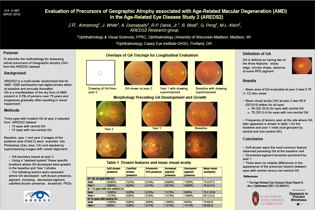
Methods: The Age Related Eye Disease Study 2 (AREDS2) is a multi-center randomized trial of 4,200 participants with stereoscopic digital color fundus photographs captured at baseline and annual visits. Thirty eyes with incident GA at year 2 visit from the AREDS2 dataset were randomly selected with 15 eyes having central GA and 15 eyes non-central GA. Using a system of stacked layers in Photoshop (San José, CA) to view specific areas in a longitudinal evaluation, we graded drusen, pigment clumping and other lesions in the baseline and year 1 visits in the specific locations where GA developed.
Results: The mean area of GA evaluated at year 2 was 0.76 (1.12) disc areas. The mean visual acuity (VA) at year 2 was 65.8(18.6) letters for all eyes, 56(20.6) for eyes with central GA and 75 (8.4) for eyes with non central GA. The frequency of the lesions seen at the site where the GA later appeared is shown in table 1 for the baseline and year 1 visits. The lesion distribution is subgrouped by central and non-central GA.
Conclusion: Soft drusen seem to be the predominant feature at the baseline visit with decreased pigment becoming prominent by year 1. There were no notable differences in the appearance of the precursor lesions between eyes with central versus non-central GA.