Comparison of Lesion Annotation Across 7-Field and Ultrawide Field Imaging in Diabetic Retinopathy (COLA)
 Cole Bacig, Nancy Barrett, Kristi Dohm, Amitha Domalpally, Barbara Blodi
Abstract
Purpose: The gold standard for diabetic retinopathy (DR) evaluation is the Early Treatment Diabetic Retinopathy Study (ETDRS) 12-step severity scale (DRSS), based on stereoscopic 7-Field (7F) color fundus photographs. Prior studies report moderate agreement between 7F and Ultra-Widefield (UWF) imaging for DR severity, with variability in lesion detection. This study compares DR lesion detection rates between paired 7F and UWF images and investigates potential causes for discrepancies.
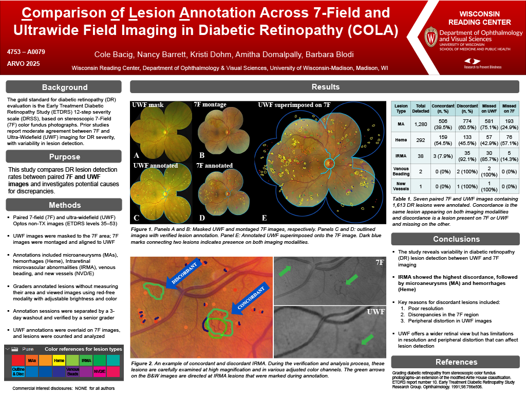
Methods: High-quality paired 7F and Optos UWF images of eyes graded ETDRS levels 35–53 were analyzed. UWF images were masked to match the 7F area using a grid, while 7F images were montaged with DualAlign’s® i2k Retina™. Lesion annotation included microaneurysms (MA), hemorrhages (Heme), intraretinal microvascular abnormalities (IRMA), venous beading, and new vessels. To minimize bias, a 3-day washout separated annotation sessions for each modality. A senior grader verified all annotations. UWF annotations were superimposed onto 7F images, aligning key landmarks (Figure 1). Lesions were categorized as concordant or discordant based on presence in both modalities, and discrepancies were analyzed.
Results: Seven paired 7F and UWF images with 1,613 DR lesions were annotated. Among 1280 detected MA, 506 (39.5%) were concordant and 774 (60.5%) discordant; of these, 581 were missed on UWF and 193 on 7F.
For 292 Heme, 159 (54.5%) were concordant and 133 (45.5%) discordant; UWF missed 57 and 7F missed 76.
Out of 38 IRMA, 3 (7.9%) were concordant and 35 (92.1%) discordant, with 30 missed on UWF and 5 on 7F. Two venous beading lesions were discordant and missed on UWF and one new vessel was discordant and missed on UWF. The top three reasons for discordant lesions were resolution differences, discrepancies in the 7F region, and peripheral distortion in UWF images.
Conclusions: This study highlights variability in DR lesion detection between UWF and 7F imaging modalities. Among the lesion types analyzed, IRMA exhibited the highest discrepancy rate, followed by MA and Heme.
While UWF imaging offers a broader field of view, its limitations in resolution and peripheral distortion affect
lesion detection which has a direct impact on DRSS.