Fundus AutoFluorescence Risk Factors for Progression of Geographic Atrophy in the Age-Related Eye Disease Study 2 (2018)
 Jeong W. Pak, Amitha Domalpally, Elivira Agron, Ronald Danis, Traci E. Clemons, Emily Y. Chew, and the AREDS2 Research Group
Abstract
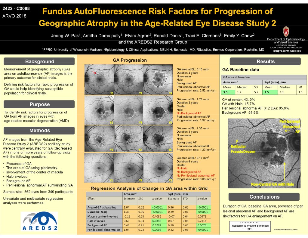
Purpose: To identify risk factors for progression of geographic atrophy (GA) from autofluoresence (AF) images in eyes with age-related macular degeneration (AMD)
Methods: AF images from the Age-Related Eye Disease Study 2 (AREDS2) ancillary study were centrally evaluated for presence and area of GA. The area of GA was measured using planimetry. In addition, area of abnormal AF surrounding GA (peri lesional) was also measured. Other AF variables evaluated include involvement of the center of macula, presence of halo and presence of background AF. Presence of GA was also confirmed on corresponding color photographs.
Results: AF images of 362 eyes from 340 participants with GA (decreased AF) with one or more years of follow-up were evaluated. The mean baseline area of GA from AF images was 3.5 mm2 (SD 5.2; median 1.2 mm2). The mean progression rate/year was 1.33 mm2 (SD 0.05) and on the square root scale 0.29 mm (SD 0.01). With multivariate analysis, baseline area of GA, duration (years from baseline), presence of background AF and peri lesional abnormal AF had significant correlation with progression rate for square root mm measurements and for the area scale (p<0.05). The presence of halo was a significant risk factor on the area scale (p=0.03) but not on the square root scale (p=0.23). Involvement of the center of macula showed no statistically significant association for GA progression.
Conclusions: Duration of GA, baseline area, presence of peri lesional abnormal AF and background AF are risk factors for GA enlargement on AF.