Multimodal Imaging Evaluation of Age-Related Macular Degeneration (AMD) in the AMD Ryan Initiative Study (ARIS) (2022)
 Andrew Dieu, Cynthia Hurtenbach, Jeong W. Pak, Tiarnan D. Keenan, Frederick L. Ferris, Charles P. Wilkinson, Christine Orndahl, Traci E. Clemons, Emily Y. Chew, Amitha Domalpally
Abstract
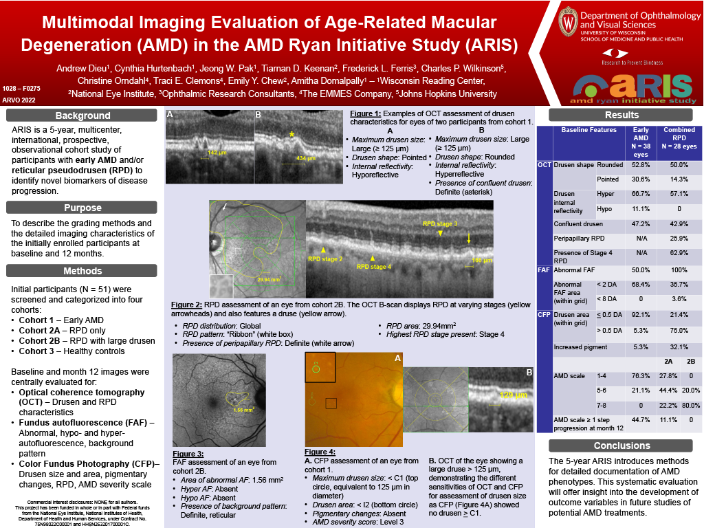
Purpose: ARIS is a 5-year, multicenter, international, prospective, observational cohort study of participants with early AMD or reticular pseudodrusen (RPD) to identify novel biomarkers of disease progression. The purpose of this abstract is to describe the detailed imaging characteristics of the initially enrolled participants.
Methods: Baseline and 12-month follow up images were evaluated for four cohorts: early AMD, RPD only, RPD and large drusen, and healthy controls. Optical coherence tomography (OCT) images were graded for drusen structure and RPD characteristics. Fundus autofluorescence (FAF) images were graded for hypo and hyperautofluorescence and background pattern. Color fundus photography (CFP) images were graded for drusen size and area, pigmentary changes, RPD, and AMD severity scale.
Results: The cohorts comprised 38 eyes with early AMD, 18 eyes with RPD only, 10 eyes with RPD and large drusen, and 36 healthy control eyes.
In baseline evaluation of the early AMD cohort, OCT showed that drusen were predominantly dome shaped (52.8%) with hyperreflective cores (66.7%). Confluent drusen were observed in 47.2% of eyes. Abnormal FAF was seen in 50% of eyes in this cohort, all with areas of abnormality less than 2 disc areas. There was no significant hypo or hyper FAF in any eyes. CFP findings showed predominantly medium drusen (92.1%) and increased pigmentation in 5.3%. There were no eyes with depigmentation. Baseline AMD severity scale was level 2–4 in 63.2% of eyes. Progression of ≥ 1 step on the AMD scale over 12 months was seen in 17 (44.7%) eyes in the early AMD cohort.
Baseline assessment of the combined RPD cohorts showed global RPD distribution in 88.9% of eyes and superior distribution in 7.4%. Peripapillary RPD was seen in 25.9%. OCT-based staging showed stage 4 RPD in 63%. Baseline AMD scale was level 6 in 32.1% eyes and level 7 in 42.9%. Progression of ≥ 1 steps on the AMD scale over 12 months was seen in 2 (7.1%) eyes in the RPD cohort, with one eye progressing to late AMD. In the RPD subset with large drusen, drusen area ≥ 1 disc area was seen in 70% of eyes at baseline, with no eyes progressing ≥ 1 step on the AMD severity scale.
Conclusions: The 5-year ARIS introduces reproducible methods for detailed documentation of AMD phenotypes. This systematic evaluation will offer insight into the development of outcome variables in future studies of potential AMD treatments.