Prevalence and Characteristics of Reticular Pseudodrusen in the Carotenoids in Age-Related Eye Disease Study 2 (CAREDS2) (2021)
 Jeong W Pak, SC Cleland, Amitha Domalpally, Zhe Liu, Barbara Blodi, S Bailey, K Gehrs, R Wallace, L Tinker, and Julie A. Mares
Abstract
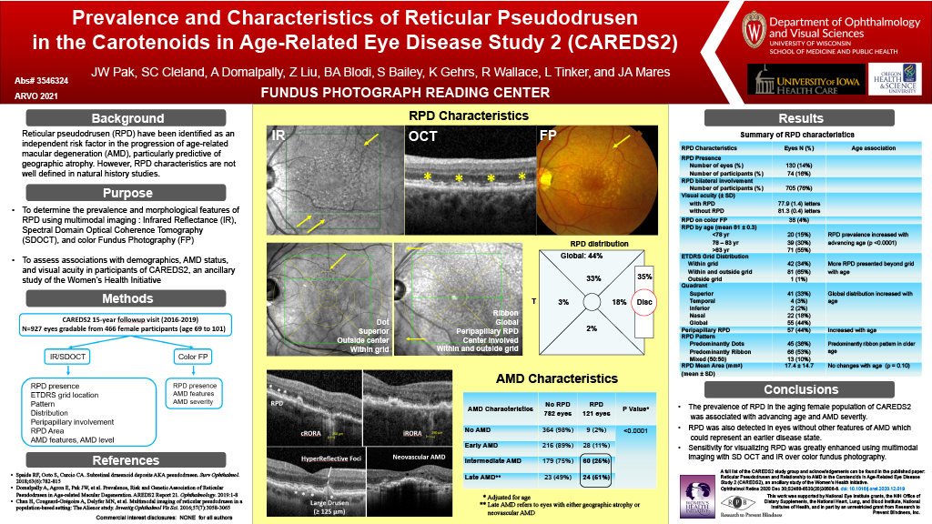
Purpose: Reticular pseudodrusen (RPD) have been identified as an independent risk factor in the progression of age-related macular degeneration (AMD), particularly predictive of geographic atrophy. However, RPD characteristics are poorly defined in natural history studies. We determined the prevalence and morphological features of RPD using multimodal imaging and assessed associations with demographics, AMD status, and visual acuity in participants of CAREDS2, an ancillary study of the Women’s Health Initiative.
Methods: Multimodal imaging included spectral domain optical coherence tomography (SD OCT) and infrared reflectance (IR) to identify RPD characteristics, such as presence, location (within or outside the ETDRS grid), peripapillary involvement, pattern, and RPD area. AMD features from SD OCT, IR, and color photographs were also assessed and AMD severity was categorized.
Results: In 927 eyes from 466 female participants (age 69 to 101), RPD were present in 130 eyes (14% of eyes, 16% of participants) and 76% participants with RPD had bilateral involvement. There was increasing prevalence with age; 7% in < 78 years, 14% in 78-83 years, and 30% in > 83 years. The AMD severity classification from the color photographs showed RPD in 2.4% of eyes with no AMD, 11.5% in early AMD, 25.1% in intermediate AMD and 51.1% in late AMD. Ribbon morphology (53%) was more common than dot morphology RPD (36%). RPD were mostly located both within and outside the ETDRS grid with primarily superior retinal distribution. Among eyes with RPD, 35% had peripapillary involvement which showed an increase with age. Mean RPD area was 17.4 (14.7) mm and was not significantly associated with age (P = 0.10) or with AMD status (P=0.60). RPD were visualized with corresponding color fundus photography in only 38 eyes (4% of total eyes). Participants with and without RPD had a visual acuity ± standard error of 77.9 (1.4) and 81.3 (0.4) letters, respectively (P = 0.02).
Conclusions: The prevalence of RPD in the aging female population of CAREDS2 was associated with advancing age and AMD severity. RPD were also detected in eyes without other features of AMD which could represent an earlier disease state. Sensitivity for visualizing RPD was greatly enhanced using multimodal imaging with SD OCT and IR over color fundus photography.