Evaluation of Diabetic Retinopathy from UltraWide Field Color Photographs (2018)
 Nancy Barrett, Kristine Lang, Amitha Domalpally, Ellie Corkery, Ralph Trane, Barbara Blodi
Abstract
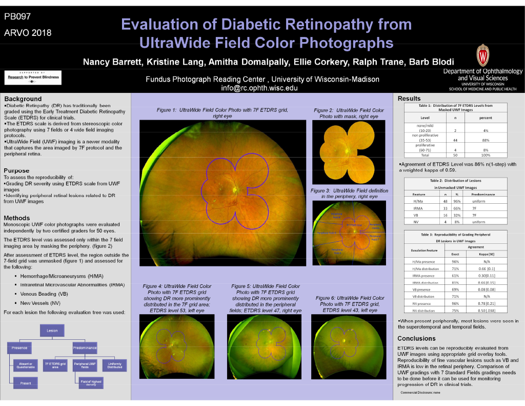
Purpose: Diabetic retinopathy (DR) has traditionally been evaluated using Early Treatment Diabetic Retinopathy Study (ETDRS) levels from stereoscopic 7 field color photographs. The UltraWide field (UWF) images (Optos, Inc) provide a view much beyond the standard 7 field images. We assessed the reproducibility of assessing ETDRS levels and DR lesions from UWF color photographs.
Methods: Monoscopic UWF color photographs were evaluated independently by two certified graders. The ETDRS level was assessed by overlaying the UWF image with a grid outlining the 7 field imaging area. The area outside the region of interest was masked. After assessment of ETDRS level, the region outside the 7 field outline grid was unmasked and assessed for presence, location and predominance of microaneurysms and hemorrhages (H/Ma), intraretinal microvascular abnormalities (IRMA), venous beading (VB) and new vessels (NV). Distribution of lesions was assessed as predominantly within 7 fields, predominantly peripheral (outside 7 fields) or uniform (equal within and outside 7 fields). Reproducibility was analyzed using percentage agreements and weighted kappa.
Results: UWF color photographs of 50 eyes were evaluated and available for comparison of gradings. There was no/mild DR in 2 (4.0%) with level < 20, non proliferative DR in 44(88.0%) with levels 35 – 53, proliferative DR in 3 (6.0%) level > 60 and one eye with scatter laser photocoagulation. Evaluation of the entire UWF image showed H/Ma in 48(96.0%) eyes, IRMA in 33 (66.0%), VB in 16 (32.0%) and NV in 4 (8.0%). IRMA and VB were predominantly within 7F, and H/MA and NV were equally distributed. When present peripherally, most lesions were seen in the superotemporal and temporal fields. The reproducibility on presence and distribution of peripheral lesions is shown in the table below.
Conclusions: ETDRS levels can be reproducibly evaluated from UWF images using appropriate grid overlay tools. Comparison of UWF gradings with 7 Standard Fields gradings needs to be before it can be used for monitoring progression of DR in clinical trials.