AI-based Multilayer OCT Segmentation Model for AMD
Rachel Linderman, Aadhi1 Balasubramanian, Madeline Pflasterer-Jennerjohn, Lucas Maakested, Jeong W. Pak, Robert Slater, Roomasa Channa, Barbara A. Blodi, Amitha Domalpally
Abstract
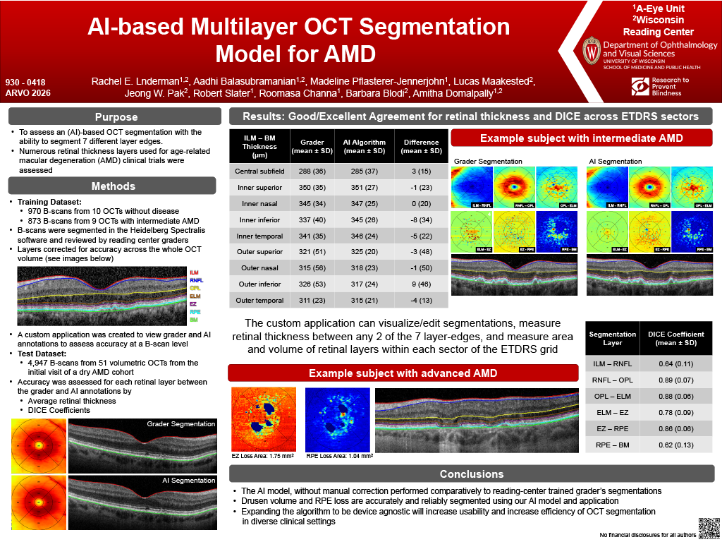
Purpose: To assess an artificial intelligence (AI)-based OCT segmentation with the ability to segment 7 different layer edges. This created 6 retinal thickness layers that are for age-related macular degeneration (AMD) clinical trials.
Methods: Using volumetric OCT scans from the WRC training library, a 2D nnSAM model was trained to segment the inner limiting membrane (ILM), bottom of the retinal nerve fiber layer (RNFL), the top of the outer plexiform layer (OPL), external limiting membrane (ELM), ellipsoid zone (EZ), retinal pigment epithelium layer (RPE), and Bruch’s membrane (BM). 970 b-scans from 10 OCTs without retinal disease and 873 b-scans from 9 OCTs with dry age-related macular degeneration (AMD) were used as the training set. All b-scans were segmented in the Heidelberg Spectralis software and the segmentation was reviewed by trained reading center graders. A custom application was created to view both the grader annotations and the AI annotations to assess for accuracy at a b-scan level. 51 OCT volumes from the screening visit from the Dark Adaptation in Participants With Age-Related Macular Degeneration clinical trial (NCT03225131) were used as the external test set. Accuracy was assessed using average retinal thickness in each of the ETDRS sectors along with DICE for each retinal layer between AI and grader annotations.
Results: Mean CST was 285µm (36.6) with AI and 288µm (36.1) with grader using ILM – BM. Across ETDRS subfields, ILM–BM thickness had minimal difference between AI and grader (-8 to 9µm). For additional thickness measurements, nerve fiber layer (ILM–RNFL) demonstrated close agreement (-4–3µm) as did inner retinal thickness (RNFL–OPL; -3–12µm). The outer retinal thickness (OPL–ELM) was consistently overestimated by AI (up to 11 µm). ELM-EZ thickness, photoreceptor thickness and RPE thickness all had near perfect correspondence. Dice scores were good to excellent for all layers (0.62–0.97).
Conclusions: This AI model, without manual correction, performed comparatively to reading-center-trained grader’s segmentations for seven retinal layer edges. Differences within each layer were minimal based on a b-scan level comparison between the AI and the grader’s segmentation. Grader intervention will provide better agreement between the segmentations. Expanding this model to be device agnostic could provide a universal and efficient method to segment multiple retinal layers requiring minimal human oversight or intervention.