AI-based Measurement of GA Area for Screening In Clinical Trials
 Mohammed Younis, Robert D. Slater, Caleb Pacheco, Sairi Zhang, Rick Voland, Rachel E. Linderman, Roomasa Channa, Amitha Domalpally
Abstract
Purpose: To develop and validate a transfer learning model for measuring geographic atrophy (GA) area using ultrawide field (UWF) fundus autofluorescence (FAF) imaging. The model was initially trained and validated on Heidelberg Spectralis images and subsequently fine-tuned for Optos UWF FAF images to expand applicability in clinical trials and increase site participation.
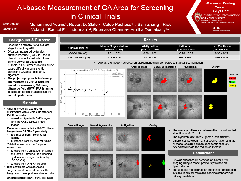
Methods: The base model utilized a Feature Pyramid Network (FPN) architecture with an EfficientNet-B5 encoder for binary class image segmentation. The model was trained on Spectralis FAF images from Age-Related Eye Disease Study 2 (AREDS2) study (601 Images from 362 eyes). Fine tuning was performed by augmenting the Spectralis trained model with a labeled data set of Optos FAF images (139 images from 139 eyes) with 16 eyes used for internal validation. Using the COCO GA dataset, the model’s performance was assessed using dice coefficient and ICC.
Results: The external dataset (n=49) had a dice coefficient of 0.804 with an ICC of 0.83. A review of the segmented annotations showed that the model had learned to limit segmentations to the macular region and not include peripheral hypoFAF lesions. In addition, the model excluded other artifacts such as lids and lashes from the segmentations.
Conclusions: Using transfer learning, a model previously trained on Spectralis FAF images was successfully adapted to detect GA on Optos UWF imaging. Expansion to additional imaging modalities enables broader participation in clinical trials and enables standardized GA segmentation. The model demonstrates the ability to generalize to different images and datasets.