AI-enabled multi-disease OCT segmentation
 Caleb Pacheco, Rachel E. Linderman, Madeline Pflasterer-Jennerjohn, Mark Banghart, Robert D. Slater, Roomasa Channa, Amitha Domalpally
Abstract
Purpose: To develop an artificial intelligence (AI) enabled OCT segmentation algorithm specifically tailored for diabetic macular edema (DME), capable of consistent performance across different OCT devices, addressing critical challenges in ophthalmic diagnostics. A secondary objective was to evaluate the performance of the model initially trained on healthy eyes when applied to DME, and to assess the impact of fine-tuning on DME-specific training data.
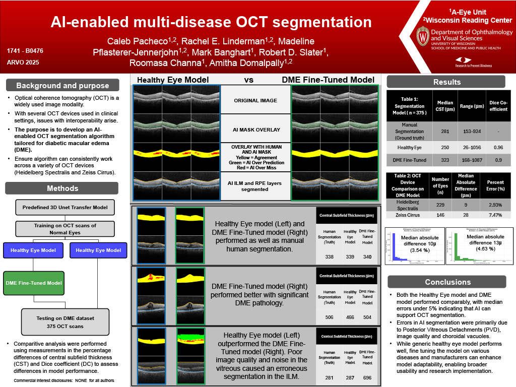
Methods: A predefined 3D Unet Transformer model was initially trained on 505 OCT volume scans of normal eyes from both Heidelberg Spectralis and Zeiss Cirrus (healthy eye model). Subsequently, the model was fine-tuned using DME-specific training data (n = x eyes). Both the original healthy eye model and the DME fine-tuned model were tested on the same DME test dataset of 375 OCT volume scans from Heidelberg Spectralis (n=229) and Zeiss Cirrus (n=146) to evaluate whether fine-tuning improved performance. Comparative analyses were performed, including differences in central subfield thickness (CST) and Dice coefficient (DC), to assess differences in performance metrics between the original and fine-tuned models.
Results: The median (range) CST for manual segmentation (ground truth) was 281 µm (153–924 µm). With the original healthy eye trained model, AI-predicted segmentation was 250 µm (26-1056 µm) with an absolute median difference of 10 µm (3.54%) and a DC of 0.96. For the DME fine-tuned model AI – prediction was 323 µm (168–1087 µm), with a median absolute difference of 13 µm (4.63%) and a DC of 0.90. Differences in the median absolute differences based on device type were observed: (Heidelberg Spectralis showed 9 µm or 2.93% and Zeiss Cirrus 28 µm or 7.47%).
Conclusions: This AI model provides a reliable device-agnostic segmentation of total retinal thickness in eyes with DME. The accuracy of predicted segmentations remained similar between the original and fine tuned models when assessing DME. Prediction errors by AI models include errors in Internal Limiting Membrane (ILM) segmentation due to Posterior Vitreous Detachment (PVD) and choroidal layers due to cystoid appearance. Exploring the performance of the healthy eye model on additional diseases will provide insights into how far AI models can be adapted to various retinal disease presentations.