Multimodal Evaluation of Macular Atrophy Associated with Neovascular AMD
Jordan Winkler, Jennifer Heathcote, Jeong W. Pak, David Lopez, Barbara A. Blodi, Amitha Domalpally
Abstract
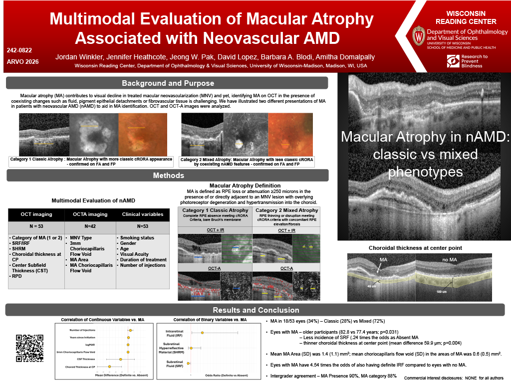
Purpose: Macular atrophy (MA) contributes to visual decline in treated macular neovascularization (MNV), yet identifying MA on OCT in the presence of coexisting changes such as fluid, pigment epithelial detachments or fibrovascular tissue is challenging. We aimed to define the characteristics of MA, describe the prevalence, atrophy phenotype, and clinical correlates of MA in a treated MNV cohort.
Methods: SD-OCT and OCT Angiography (OCTA) of 45 patients (53 eyes) with nAMD undergoing anti-VEGF treatment were included. An experienced grader assessed the presence of MA and other morphological features with a multimodal approach; Clinical variables were also recorded. On OCT, MA is defined as RPE loss or attenuation >250 microns in the presence of or directly adjacent to an MNV lesion with overlying photoreceptor degeneration and hypertransmission into the choroid. MA was further categorized as 1) complete RPE absence meeting cRORA criteria with a bare or mostly bare Bruch’s Membrane similar to typical GA, and 2) RPE thinning or disruption meeting cRORA criteria with concomitant RPE elevation and/or fibrosis.
Results: MA was present in 17/53 eyes (32%) with cRORA-like presentation (category 1) in 5(29%) and with fibrosis/ RPE elevation (category 2) in 12 (71%). Eyes with MA were from older participants (82.8 vs 77.4 years; p=0.010), had worse vision (log MAR difference −0.171; p=0.038) and thinner choroid (mean difference 71.7 μm; p≈0.002). IRF was also associated with MA (OR 3.61; p=0.038) and all eyes with atrophy had EZ loss. Other variables such as gender, smoking, duration of treatment, number of injections, and OCT features such as CSF thickness, MNV type, SHRM and SRF showed no significant differences. Intergrader agreement on presence of MA and MA subtyping was 90% and 88%, respectively.
Conclusions: Choroidal thinning showed the strongest structural association with MA. The two MA subtypes demonstrated distinct OCT phenotypes. Longitudinal data are needed to understand their trajectories and visual impact.