Features Associated with Geographic Atrophy from Autofluorescence Images (2012)
 Amitha Domalpally, Ronald P Danis, Ashwini Narkar, Barbara A. Blodi, Emily Chew, Traci Clemons
Abstract
Purpose: To evaluate the features associated with progression of Geographic Atrophy (GA) from Autofluorescence (AF) images.
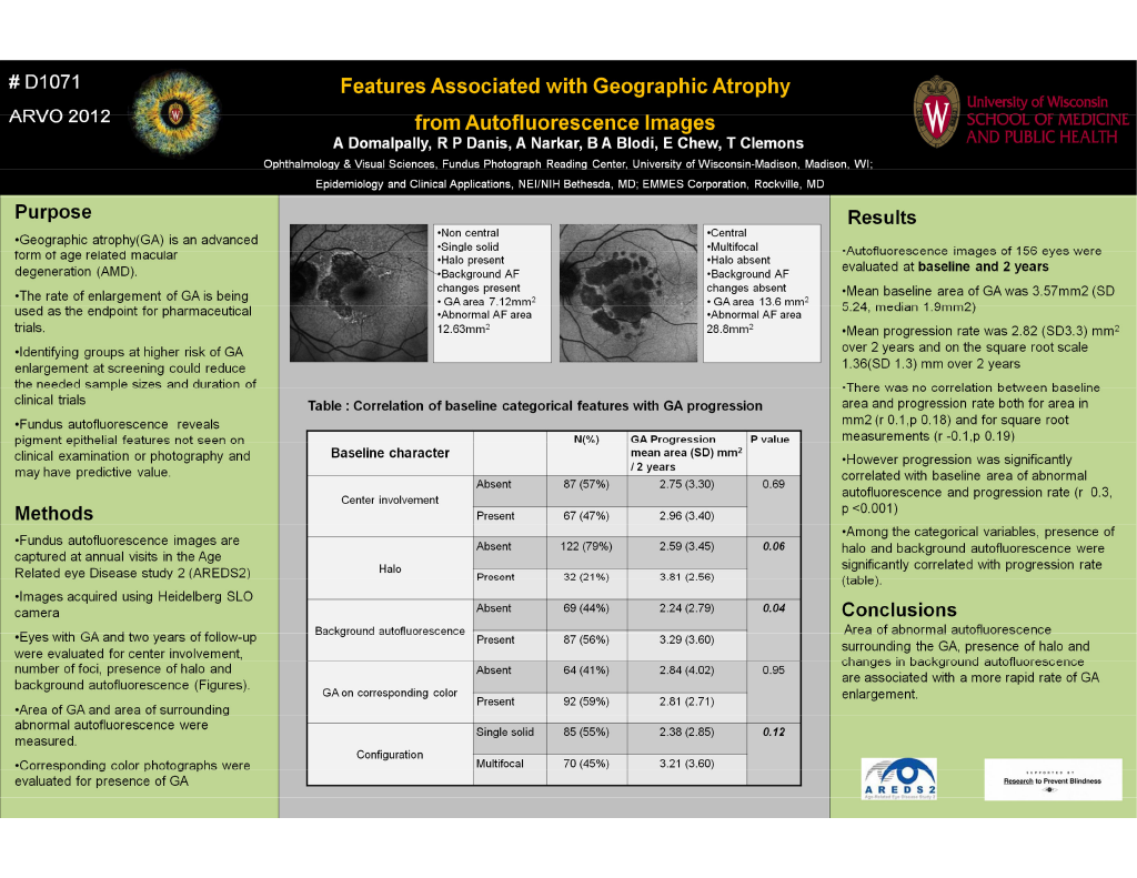
Methods: AF images (from scanning laser ophthalmoscopy) images are captured at annual visits in the Age Related Eye Disease Study 2 (AREDS2) ancillary study. Eyes with GA and two years of follow-up were evaluated for center involvement, configuration (unifocal or multifocal),presence of halo and background AF(changes outside the grid; see figure). The area of GA and the area of AF abnormalities surrounding GA within the grid (as shown by the dotted lines in the figure) were measured using planimetry. The presence of GA on corresponding color photos
was also evaluated.
Results: AF images of 156 eyes with GA at baseline and two year follow-up were evaluated. The mean baseline area of GA from AF images was 3.57 mm2 (SD 5.24; median 1.19 mm2), with 54% of lesions multifocal. The mean progression rate/year was 1.41mm2 (SD 1.65) and on the square root scale 0.68mm (SD 0.65). The baseline measurements did not correlate with progression rate both with area (r 0.1, p0.18) and square root measurements (r-0.1, p0.19). The baseline area of abnormal AF surrounding GA correlated with progression rate (r = 0.3, p<0.001). Of the categorical features evaluated, the presence of halo (p=0.06) and presence of background AF changes (p=0.04) had significant correlation with progression rate.
Conclusions: Area of abnormal AF surrounding the GA, presence of halo and changes in background AF are associated with a more rapid progression of GA on AF.