Characterization of OCT biomarkers and endpoints in intermediate AMD
 Jordan Winkler, Jennifer Heathcote, Jeong W. Pak, Rick Voland, Barbara A. Blodi, Amitha Domalpally
Abstract
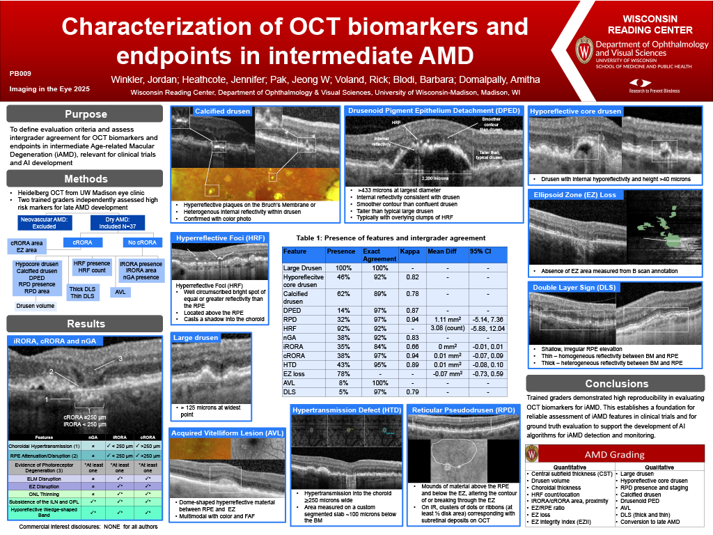
Purpose: To define evaluation criteria and intergrader agreement for assessing intermediate Age-related Macular Degeneration (iAMD) optical coherence tomography (OCT) biomarkers and endpoints in eyes with nonexudative AMD, relevant for clinical trials and AI development.
Methods: Trained graders independently evaluated Heidelberg OCT volume scans (20°x20°, 97 B scans) in eyes with intermediate AMD enrolled in the AREDS2 study at UW Madison. Graders assessed the presence of high risk markers: large drusen, hyporeflective core drusen, calcified drusen, drusenoid PED (DPED), reticular pseudodrusen (RPD), hyperreflective foci (HRF), nascent geographic atrophy (nGA), incomplete retinal pigment epithelium and outer retinal atrophy (iRORA), and outcomes including complete retinal pigment epithelium and outer retinal atrophy (cRORA), hypertransmission defect (HTD), Acquired Vitelliform Lesion (AVL) and double layer sign (DLS). Graders also assessed the area of iRORA, cRORA, HTD, ellipsoid zone (EZ) loss, and RPD, as well as HRF count. Each feature was assessed using criteria based on the published literature on iAMD biomarkers. Reproducibility of presence was analyzed as exact agreement with kappa value and area was compared by Intraclass correlation (ICC).
Results: Of 37 eyes from 28 participants, large drusen were present in all eyes (100%) with hyporeflective core drusen in 17 (46%), calcified drusen in 23 (62%), DPED in 5 (14%), RPD in 12 (32%) with mean area (SD, mm2) as 4.05 (10.3), HRF in 34 (92%) with mean count (SD) as 7.24 (8.98), nGA in 14 (38%), iRORA in 13 (35%) with mean area (SD) as 0.012 (0.022), cRORA in 14 (38%) with mean area (SD) as 0.173 (0.407), HTD in 16 (43%) with mean area (SD) as 0.232 (0.508), AVL in 3 (8%), DLS in 2 (5%), and mean area of EZ loss (SD) was 0.841 (1.164). Table 1 shows the intergrader agreement on these OCT biomarkers.
Conclusions: Trained graders demonstrated high reproducibility in evaluating OCT biomarkers for iAMD. This establishes a foundation for reliable assessment of iAMD features in clinical trials and for ground truth evaluation to support the development of AI algorithms for iAMD detection and monitoring.