Comparison of Standard 7-Field, Clarus, and Optos Ultra-Widefield Imaging Systems for Diabetic Retinopathy Assessment (2023)
 Nancy Barrett, Nicole Duncan, Christian Kim, Sheila Watson, Andy Ewen, Lauren Kroth, Rick Voland, Amitha Domalpally, Barbara Blodi
Abstract
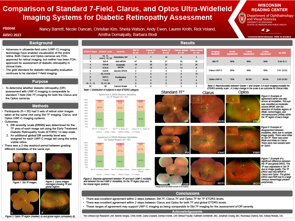
Purpose: Advances in ultrawide-field color (UWF-C) imaging technology have enabled visualization of the entire retina. This study aims to determine whether diabetic retinopathy (DR) assessment with UWF-C imaging is comparable with the current gold standard, standard 7-field (Std 7F) imaging for both the Clarus and the Optos cameras.
Methods: Participants had retinal color images taken using Std 7F imaging, Clarus, and Optos UWF-C imaging systems. DR severity scale (DRSS) was determined for the 7F area of each image set using the Early Treatment Diabetic Retinopathy Scale (ETDRS) 12-step scale. An additional global DR severity level was assigned for each UWF-C image set using the entire visible retina. Two graders assessed all images independently with adjudication by a third grader when agreement was not exact. There was a washout period of two days between grading each modality for the same patient.
Results: 50 eyes from 30 participants were evaluated. The 7F DR level was ungradable for 2 (4.0%) Std 7F image sets, 1 (2.0%) Clarus images, and 0 (0.0%) Optos images. Global DR level could not be assessed in 1 (2.0%) Clarus image set and 1 (2.0%) Optos image set. Comparing 7F DR levels between Std 7F and Clarus, agreement was exact in 55.0% of the eyes, within one step in 93.6% of the eyes, and within 2 steps in 100% of the eyes (wĸ = 0.74). Comparing 7F DR levels between Std 7F and Optos, the results were 54.0%, 81.2%, and 89.6% respectively (wĸ = 0.56). The results of Clarus Global relative to Optos Global were 56.0%, 87.5%, and 93.8% (wĸ = 0.68) respectively. In eyes identified to have PDR on Stad 7F, disagreement was found in 1 eye with Clarus and 7 eyes with Optos imaging. Intergrader agreement for 7F ETDRS level was within 2 steps 100.0% of the time for 7F, 97.8% of the time for Clarus, and 92.0% of the time for Optos.
Conclusion: When comparing the DR levels determined within the 7F and global areas, our study found exact agreement between Std 7F and both UWF-C modalities in about half of all images. The majority of all eyes showed agreement within 1 step. These ranges of agreement may support UWF-C imaging as being comparable to Std 7F imaging for the assessment of DR severity.