Reading Center Measurement of Capillary Non-Perfusion Using Ultrawidefield (UWF) Fluorecein Angiography in Two Prospective Clinical Trials of Retinal Vein Occlusion and Diabetic Retinopathy (2017)
 Ellie Corkery, Amitha Domalpally, Ruth Shaw, Kelly Warren, Sheila Watson, Barbara Blodi
Abstract
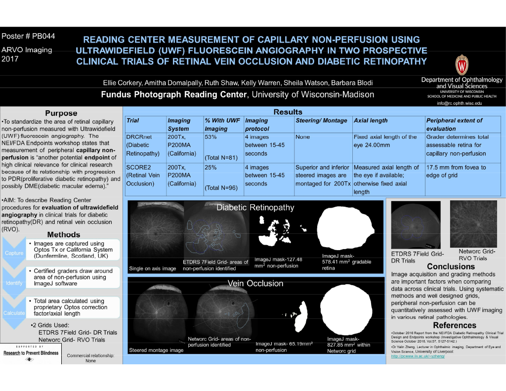
Purpose: According to the October 2016 Report from the NEI/FDA Diabetic Retinopathy Clinical Trial Design and Endpoints workshop (Investigative Ophthalmology & Visual Science October 2016, Vol.57, 5127-5142.), “Change in retinal capillary nonperfusion area over time as measured with UWF fluorescein angiography to quantitatively measure peripheral capillary nonperfusion is another potential endpoint of high clinical relevance for clinical research because of its relationship with progression to PDR(proiferative diabetic retinopathy) and possibly DME(diabetic macular edema).” At the Fundus Photograph Reading Center, UWF angiography is being evaluated as part of several large clinical trials. The objective of this submission is to present current reading center methodologies for evaluating retinal non perfusion in UWF angiography of eyes with vein occlusion and diabetic retinopathy in clinical trials.
Methods: All UWF images were captured on the Optos 200 Tx or California systems (Dunfermline, Scotland, UK). Certified evaluators identified capillary nonperfusion, utilizing Image J software to draw around the areas of non-perfusion; total area was calculated by applying a proprietary Optos correction factor. The table depicts the similarities and differences between two clinical trials evaluating capillary nonperfusion imaged with UWF angiograms at the initial visit.
Results: In the diabetic trial, no images were montaged and a standard axial length measurement (24.00 mm) was used to perform the area calculation. The ETDRS (Early Treatment Diabetic Retinopathy Study) 7 field grid outline was used and nonperfusion was measured both within and beyond the outline; extent measured to the outer border of visible retinal vasculature. In the vein occlusion trial, 63 of the 96 subjects had images that were steered and available for montage. Area of non-perfusion was assessed within the central macular grid and all 12 subfields of the Networc grid. The outer border of the Networc grid used in this trial has a radius of 17.5mm from the foveal center.
Conclusions: Image acquisition and grading methods are important factors when comparing data across clinical trials. Using systematic methods and well designed grids, peripheral non perfusion can be quantitatively assessed with UWF imaging in a variety of retinal diseases.