Illustration of the AMD Severity Scale from the Age-Related Eye Diseases Study (2004)
 Jane Armstrong, Matthew D. Davis, R.E. Gangnon, L. –Y. Lee, Ronald Klein, Barbara E. Klein, R. C. Milton, F.L. Ferris, Larry D. Hubbard
Abstract
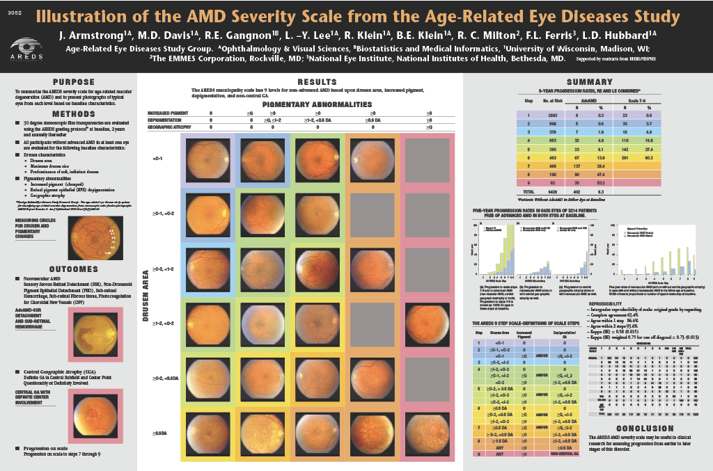
Purpose: To summarize the AREDS severity scale for age–related macular degeneration (AMD) and to present photographs of typical eyes from each level.
Methods: AREDS enrolled persons without advanced AMD in at least one eye and followed them for progression to advanced AMD – neovascular AMD or central geographic atrophy (GA). Baseline color photos were graded for drusen characteristics (size, area, type), pigment abnormalities (increased retinal pigment, RPE depigmentation), and non–central GA, and annual follow–up photos were graded for progression. Clinical judgment and regression analyses were used to construct an AMD severity scale, organized by observed predictive value of various baseline features for later onset of advanced AMD or more severe non–advanced AMD. Various subsets were analyzed; results below are from 6426 eyes of 3214 persons without advanced AMD in either eye at baseline and with gradings of the 5–year follow–up visit available as of May, 2001.
Results: The AREDS scale has 9 levels for non–advanced AMD based upon drusen area, increased pigment, and depigmentation/non–central GA. It also has 2 levels for advanced AMD – central GA and neovascular AMD. Among the 9 levels for non–advanced AMD, 5–year progression rates to advanced AMD were: level 1 – 0.3%, 2 – 0.6%, 3 – 1.9%, 4 – 4.9%, 5 – 6.1%, 6 – 13.9%, 7 – 28.1%, 8 – 47.4%, and 9 – 53.2%. Morphologic criteria defining each level and typical examples from each will be shown.
Conclusions: Researchers desiring to use the AREDS AMD severity scale for clinical and epidemiologic studies would need to become familiar with the criteria for and typical appearance of eyes in each level.