Imaging Features of Intraretinal Microvascular Abnormalities in Diabetic Retinopathy (2023)
 Christian Kim, Jeong W. Pak, Nancy Barrett, Nicole Duncan, Mary K. Wilda, Kristi Dohm, Jen Heathcote, Barbara. A. Blodi, Rick Voland, Amitha Domalpally
Abstract
Purpose: To compare characteristics of intraretinal microvascular abnormalities (IRMA) on color fundusphotography (CFP) to UWF-FA. We hypothesize that IRMA on CFP have similar characteristics tocorresponding lesions seen on UWF-FA.
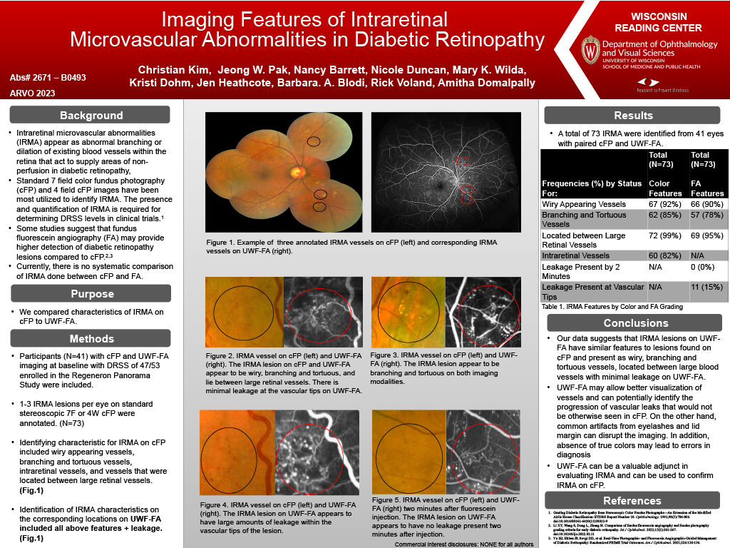
Methods: Participants with ultrawide field imaging at baseline and a DR Severity Scale of 47/53 (moderateand moderately severe NPDR), enrolled in the Regeneron Study were included. The location of up to threeIRMA patches per eye were annotated on standard 7 field and 4 field CFP and the characteristics documentedincluded wiry appearing vessels, branching and tortuous vessels, low-lying (intraretinal) vessels when viewedunder stereoscope imaging, and vessels that were located between large retinal vessels. Wiry appearingvessels are defined as vessels small in diameter, while branching and tortuous vessels contained varioustwists and turns. Corresponding locations were examined on UWF-FA and the same characteristics weredocumented, and with an added characteristic of vessels containing leakage within vascular tips (Fig. 1).
Results: A total of 63 IRMA were identified from 30 eyes with paired CFP and UWF-FA. On CFP, 92% of IRMAlesions showed wiry appearing vessels, 86% showed branching and tortuous vessels, 92% of vesselsappeared to be low lying vessels when viewed under stereoscope imaging, and 100% of IRMA were locatedbetween large retinal vessels. On corresponding UWF-FA, 91% of lesions showed wiry appearing vessels,79% showed branching and tortuous vessels, 70% of vessels did not have leakage present at vascular tips,and 95% of IRMA were located between large retinal vessels. When comparing exact lesions between imagingtechnologies, 94% of wiry appearing vessels, and 89% of branching and tortuous vessels were in exactagreement with each other.
Conclusions: The current gold standard for identifying IRMA is through CFP. Our data suggests that IRMAlesions on UWF-FA have similar features to lesions found on CFP and present as wiry, branching and tortuousvessels, located between large blood vessels with minimal leakage on UWF-FA. Although CFP isrecommended for identification of IRMA lesions, it has limitations such as image quality being dependent onpupil diameter, and its susceptibility to opacities of the media. UWF-FA can be a valuable adjunct in evaluating IRMA and can be used to confirm IRMA on CFP.