Optical Coherence Tomography in the Evaluation of Neovascular Lesions and Serous Pigment Epithelial Detachments in Polypoidal Choroidal Vasculopathy (2016)
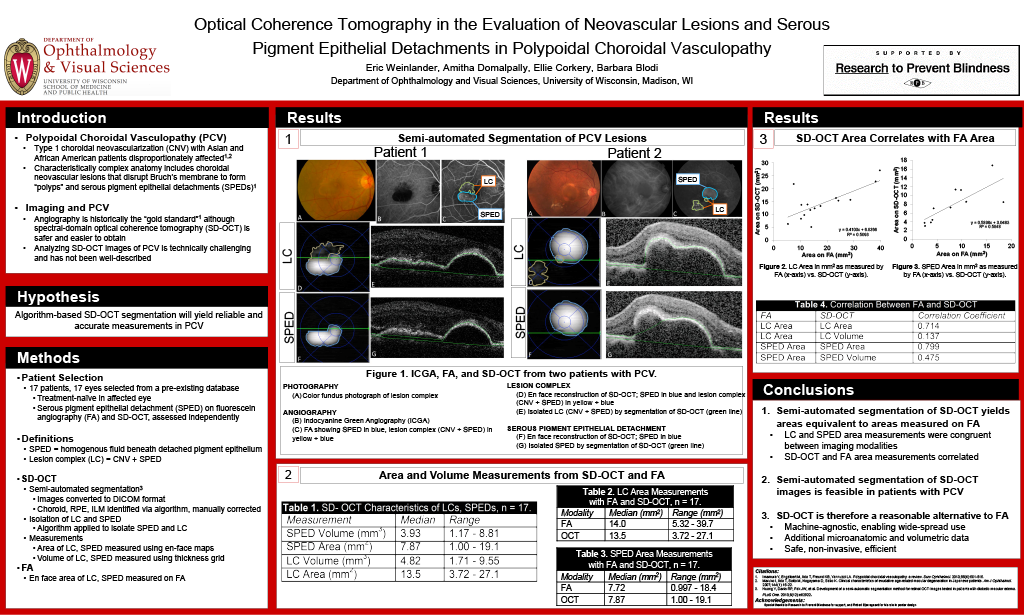 Eric Weinlander, Amitha Domalpally, Ellie Corkery, Barbara Blodi
Abstract
Purpose: Spectral domain optical coherence tomography (SD-OCT) has not routinely been used to measure features of polypoidal choroidal vasculopathy (PCV) due to its anatomic complexity. We hypothesize that a novel algorithm-based semi-automated approach to grading SD-OCT will overcome this difficulty to yield reliable and accurate measurements in PCV.
Methods: 17 patients with untreated PCV with serous pigment epithelial detachment (SPED) detected both on Fluorescein Angiography (FA) and SD-OCT were selected from a pre-existing database. SD-OCT images from various manufacturers were converted to a DICOM format, and the segmentation algorithm was applied to quantify the lesion complex (LC; neovascular lesion and associated SPED; figure 1A). The SPED was also isolated and segmented using the same method (figure 1B). OCT thickness grid with volume measurements were used to quantify the LC and SPED. En face maps were generated to measure area of LC and SPED. The lesion area and volumes obtained from FA and SD-OCT were correlated using the Pearson coefficient.
Results: Median area of LC on SD-OCT and FA was 14.0mm2 and 13.5mm2, respectively and median area of SPED on SD-OCT and FA was 7.72mm2 and 7.87mm2. Area measurements by SD-OCT and FA correlated well; r = 0.714 for LC and r = 0.799 for SPED. Median SD-OCT volume for LC was 4.82 mm3 and for SPED was 3.93 mm3 . Volume measurements by SD-OCT did not correlate with any of the FA area measurements. Volume of LC did not correlate with corresponding enface area but volume of SPED did correlate with corresponding enface area( r XX). , (r = 0.730).
Conclusions: Area measurements of LC and SPED by SD-OCT and FA are comparable. Thus, quantitative assessment of PCV using SD-OCT provides volume and area measurements that can be used to monitor anatomical outcomes in clinical trials. Longitudinal followup to quantify neovascular lesion regression and correlation with visual acuity would provide insight into the management of PCV.