Evaluation of Lens Opacities Using Fundus Reflex Photographs in the Age-Related Eye Disease Study 2 (AREDS2) (2013)
 Erika Treichel, Amitha Domalpally, Ronald Danis, Susan Reed, Ashwini Narkar, Traci Clemons, Emily Chew, and the AREDS2 Research Group
Abstracts
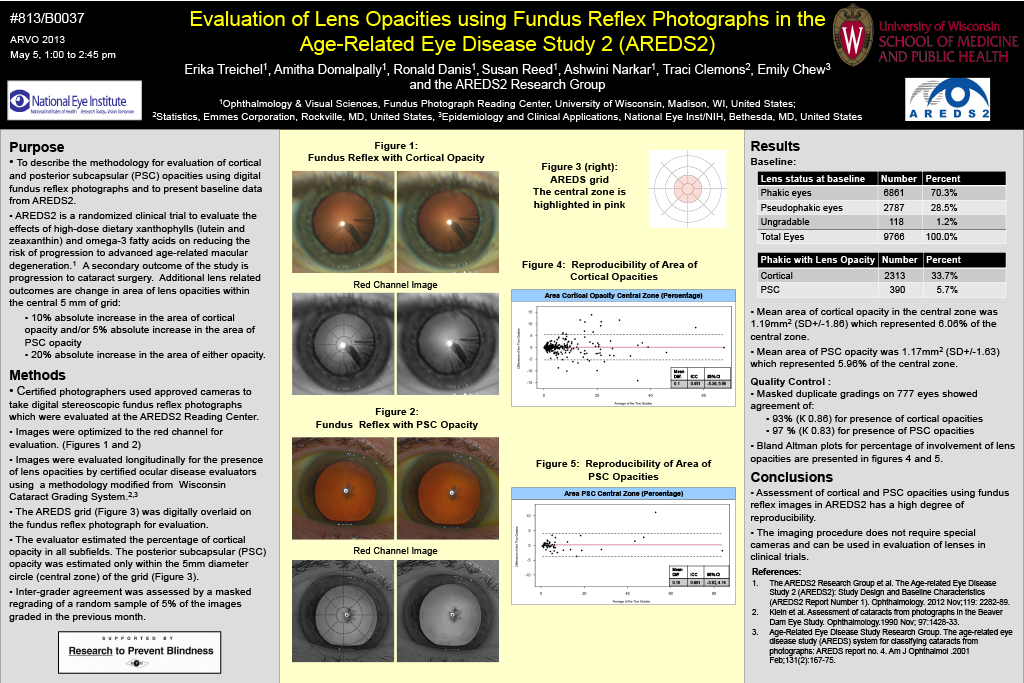
Purpose: To describe the methodology for evaluation of cortical and posterior subcapsular cataracts using digital fundus reflex photographs and the baseline lens data in AREDS2. The goal of AREDS2 is to study the effects of high-dose dietary xanthophylls (lutein and zeaxanthin) and omega-3 antioxidants on development and progression of cataract.
Methods: Digital stereoscopic fundus reflex photographs were taken by certified photographers using approved cameras for evaluation at the AREDS2 Reading Center. Photographs were evaluated longitudinally for the presence of lens opacities by certified ocular disease evaluators using standardized procedures. A grid with 17 subfields divided by 4 concentric circles and 8 equally-spaced lines was digitally overlaid on the fundus reflex photograph for evaluation. The evaluator estimated the percentage of cortical opacity in all subfields and posterior subcapsular (PSC) opacity in the central zone. Inter-grader agreement was assessed by regrading a random sample of 5% of the images graded in the previous month.
Results: At baseline 9766 fundus reflex images were evaluated from 4883 subjects. Of these, 6861 (70.3%) were phakic, 2787 (28.5%) were pseudophakic, and 118 images (1.2%) could not be evaluated. Of the phakic eyes, 2313 (33.7%) had cortical and 390 (5.7 %) had PSC opacities. Mean area of cortical was 5.01mm2 (SD+/-6.3) which represented 14.12% of the grid. Mean area of PSC was 1.17mm2 (SD+/-1.63) which represented 6.15% of the central zone. Masked duplicate gradings on 777 eyes showed an agreement of 93% (К 0.86) for presence of cortical opacities and 97 % (К 0.83) for PSC. The mean difference between the two grades was -0.03mm2 (95% CI: -5.31, 5.25) for cortical opacity and 0.03mm2 (95% CI: -0.75, 0.81) for PSC.
Conclusions: Assessment of cortical and PSC opacities using fundus reflex images in AREDS2 had a very high degree of reproducibility and this methodology can be used in evaluation of lenses in clinical trials.