Association of Outer Retinal Layer Morphology with Visual Acuity in Patients with Macular Edema Secondary to Retinal Vein Occlusion – A SCORE Study Ancillary Study (2010)
 Amitha Domalpally, Qian Daisy Peng, Ronald P Danis, Barbara Blodi, Ingrid U Scott, Michael Ip, and SCORE Study Research Group
Abstract
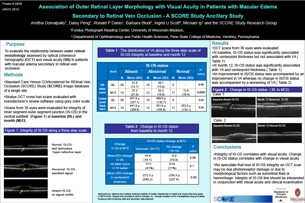
Purpose: To evaluate the relationship between outer retinal morphology assessed by optical coherence tomography (OCT) and visual acuity (VA) in patients with macular edema secondary to retinal vein occlusion.
Methods: High resolution cross hair scans captured with time domain OCT (Stratus) (512 A-scans per B-scan) available from the SCORE Study image database of a single site were evaluated for outer retinal morphology features at baseline and months 4, 8, and 12. The integrity of the inner segment outer segment junction (IS-OS) was assessed on a 3 step scale: absent, questionable, and present. Photoreceptor length was measured from the outer plexiform layer to the retinal pigment epithelium using software calipers. Best corrected ETDRS VA was available for all participants.
Results: OCT scans of 36 eyes were evaluated for outer retinal morphology. There was no difference in VA in eyes with absent (n= 26) or abnormal (n= 10) IS-OS at baseline (51.8 vs. 55.7 p = 0.46). Baseline center point thickness (CPT) was significantly different between these groups with a mean of 657 µ in eyes with absent IS-OS and 469 µ in eyes with abnormal IS-OS (p = 0.02). At month 12, the IS-OS was absent in 13, abnormal in 12 and normal in 11 eyes. Mean VA was 37.2, 52.3 and 72.6, respectively (p<0.001). CPT was also significantly different among eyes with varying IS-OS integrity (p=0.04). Among eyes that improved in IS-OS status (n=17) from baseline to month 12, the mean change in VA was +6.4 letters with 7 (41%) eyes gaining ≥ 15 letters. Among eyes that showed no change in IS-OS status (n=18) between baseline and month 12, the mean change in VA was – 5.9 letters with only 3 (16%) eyes gaining ≥ 15 letters. The results were similar when analyzed separately for central and branch retinal vein occlusion subgroups.
Conclusions: The integrity of the IS-OS can be interpreted from high resolution cross hair scans from time domain OCT. Status of IS-OS correlates with VA at follow up, and change in IS-OS status correlates with change in VA, in eyes with retinal vein occlusion enrolled in the SCORE Study.